Home > E. Pathology by systems > Locomotory system > Bones > giant cell reparative granuloma
giant cell reparative granuloma
Tuesday 4 April 2006
Giant Cell Reparative Granuloma; GCRG; giant cell granuloma of bones; giant cell granuloma of bones, giant-cell reparative granuloma, giant-cell reparative granuloma (GCRG) ; giant cell reaction of bones;
| WP | PO |
Digital slides
HPC:86 : Peripheral giant cell reparative granuloma of bones
HPC:177 : Peripheral giant cell reparative granuloma of bones
Images
WP http://www.webpathology.com/case.asp?case=662
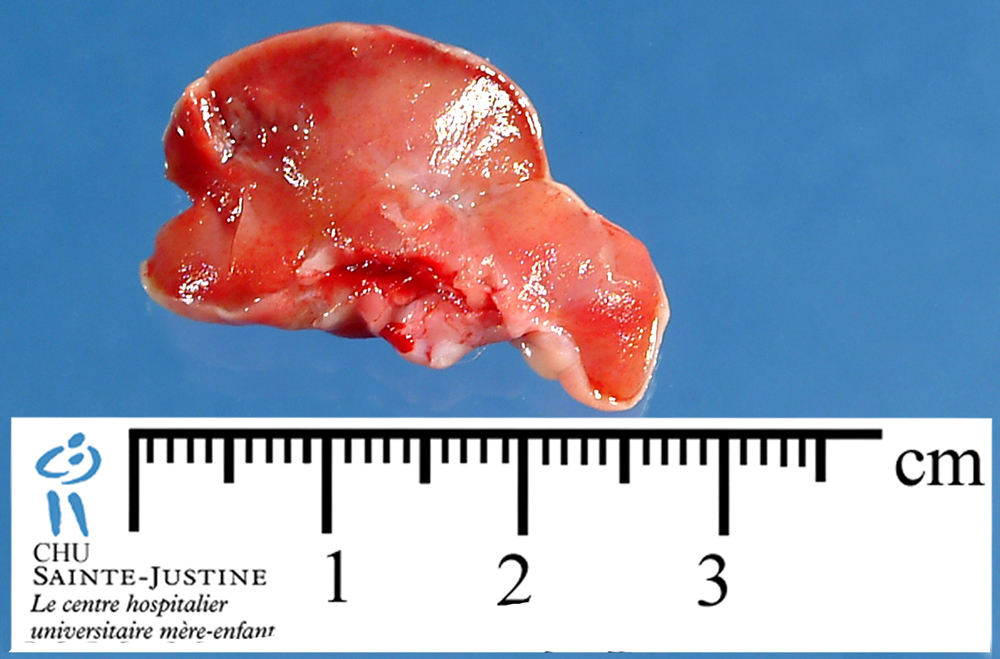
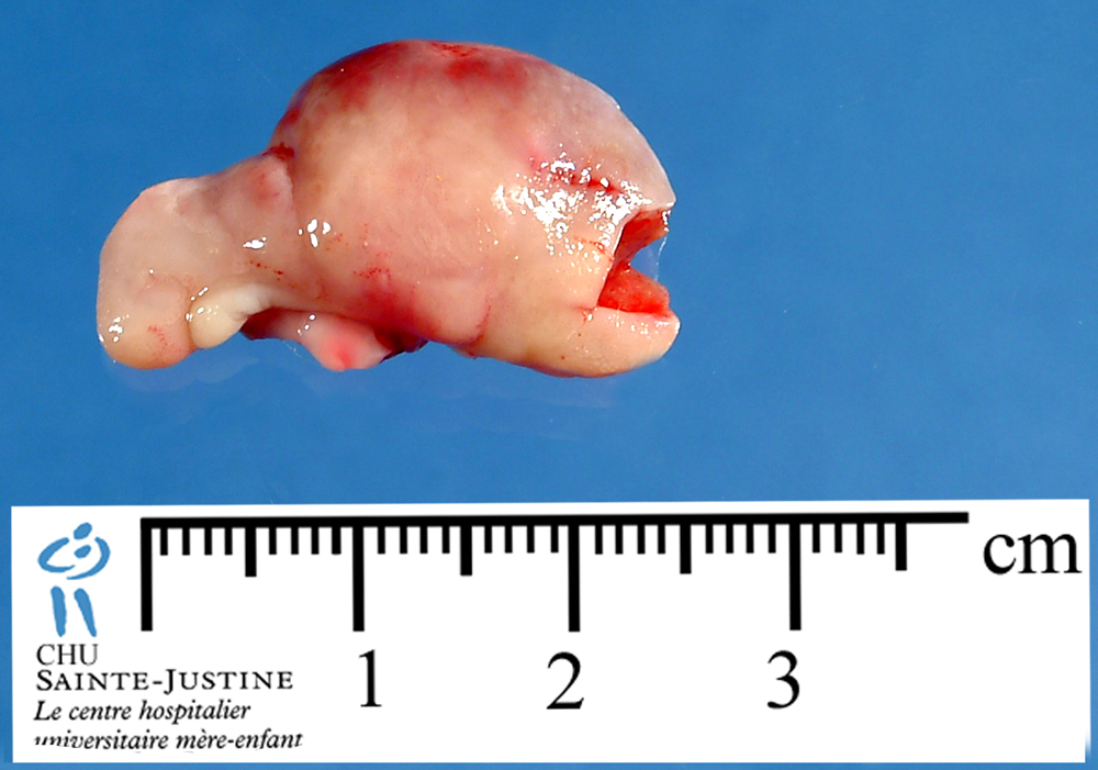
Definition: Giant-cell reparative granuloma (GCRG) is an uncommon benign reactive intraosseous lesion. GCRG occurs in the skull, jaw, hand, foot, and facial bones and rarely in other skeletal sites.
GCRG is a solitary, lytic, expanded lesion and infrequently may extend into the surrounding soft tissue.
This solitary lesion has a predilection for the bones of the hands and feet, facial bones, skull, and jaw. It is a non-neoplastic, reactive lesion of unknown etiology. It may be due to injury, trauma, or intraosseous hemmorhage.
Nota bene: The name "solid variant of aneurysmal bone cyst" has also be given to this tumor but is a different entity and must be avoided.
Synopsis
This solitary lesion has a predilection for the bones of the hands and feet, facial bones, skull, and jaw.
It is a non-neoplastic, reactive lesion of unknown etiology.
All ages are affected.
Pain and swelling are common symptoms. There is usually no distinct mass.
The lesion is lytic, actively destroying bone, it may thin or destroy the nearby cortex, but no perisosteal reaction is seen.
Intralesional curettage and bone grafting are sufficient.
Epidemiology
The age range is from childhood to older adults. There does not appear to be a definite age of peak incidence for this lesion.
Most cases are in the jaw, facial bones, and skull, short bones of the hands and feet, and a few cases have been reported in the long bones of the upper and lower extremities.
Symptoms and Presentation
Pain and swelling, or a slight pathological fracture
Clinical differential Diagnosis: ABC, GCT, brown tumor, metastasis, infection. The appearance of the lesion may be aggressive.
Radiology
The lesion is lytic, slightly expansile, actively destroying bone, it may thin or destroy the nearby cortex, but no perisosteal reaction is seen.
The lesion does not seem capable of forming an extraosseous mass.
The borders may be indistinct, and the lesion may have a slightly aggressive appearance. The location within the bone is variable, and may even be subperiosteal.
In the author’s series, no overlying thin layer of reactive bone is seen covering the extraosseous portion of the tumor, which may be present in GCT and ABC’s.
There is no mineralization of the matrix.
These lesions cannot be readily distinguished from other active, lytic processes such as ABC, GCT, or primary sarcoma, or metastatic cancer.
Microscopy
Histologically, it is composed of fibrous stroma with spindle-shaped fibroblasts, multinucleated giant cells, and inflammatory mononuclear cells. Areas of hemorrhage are uniformly present.
There is reactive granulation tissue, spindle-shaped fibroblasts, focal hemorrhage, and scattered small giant cells that have few nuclei. The giant cells may cluster around areas of hemorrhage. Irregular bony trabeculae have osteoclastic giant cells on some surfaces. Some cases have had much more frequent giant cells.
Types
central giant cell reparative granuloma (maxillary, mandible) (central GCRG)
peripheral giant cell reparative granuloma (non jaw GCGB or peripheral GCRG)
Differential diagnosis
giant cell-rich lesions of bone :
- aneurysmal bone cyst (and "solid aneurysmal bone cyst")
- giant cell tumor of bone
- brown tumor of hyperparathyroidism
- non-ossifying fibroma (if storiform pattern of spndle cells and foam cells)
GCRG may be difficult to distinguish this entity from an aneurysmal bone cyst, giant-cell tumor, or brown tumor of hyperparathyroidism because of roentgenographic and histologic similarities.
The presence of giant cells leads to the potential for confusion with ABC, GCT, PVNS, and brown tumor.
Immunochemistry
alpha1-antichymotrypsin +
alpha1-antitrypsin +
factor XIIIa +
stromal cells: vimentin+, smooth muscle actin + (SMA +)
osteoclast-like cells: CD68+, vimentin+, leucocyte common antigen (LCA+)
Associations
Noonan syndrome (9660063, 16860226) (with CGCG)
Cytogenetics
Management
Accurate diagnosis is essential for appropriate treatment.
No diagnostic lab findings exist, but labs may be useful to eliminate other tumors in the differential.
Serum calcium, phosphorus, and parathyroid hormone levels should be measured.
Curettage and bone graft are effective treatments for both primary lesions and recurrences.
Curettage, with bone graft or other bone void filler to treat the bone defect.
Preferred Margin for this Tumor: intralesional.
Prognosis
Second recurrences are rare.
Links
See also
giant cell-rich lesions of bones
References
Wold LE, Dobyns JH, Swee RG, Dahlin DC. Giant cell reaction (giant cell reparative granuloma) of the small bones of the hands and feet. Am J Surg Pathol. 1986 Jul;10(7):491-6. PMID: 3728789
Lorenzo JC, Dorfman HD. Giant-cell reparative granuloma of short tubular bones of the hands and feet. Am J Surg Pathol. 1980 Dec;4(6):551-63. PMID: 7212148
Hirschl S, Katz A. Giant cell reparative granuloma outside the jaw bone. Diagnostic criteria and review of the literature with the first case described in the temporal bone. Hum Pathol. 1974 Mar;5(2):171-81. PMID: 4590573
Giant-cell reparative granuloma of the hand and foot bones Ratner-V; Dorfman-HD Department of Orthopaedic Surgery, Montefiore Medical Center, Bronx, New York 10467-2490. Clin-Orthop. 1990 Nov(260): 251-8